EBit30
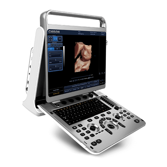
Display Unit
15" High Resolution LED Monitor
500 GB HDD and 4 USB
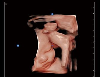
Super Image Module
Multiple Compound Imaging, Speckle Reduction Algorithms, FHI, X-Contrast, Q-Beam, Q-Flow, Q-Image.
Analysis
Measurement & Calculation software packages: General OB&GYN, Cardiac
Modes
B, 2B, 4B, B/M, B/BC, CFM, PW, Power Doppler, Directional PD, Instant Triplex, Duplex, Trapezoidal, Chroma B&M&PW
9 Breakthrough Technologies
- Space Compound Imaging – Traditional Compound imaging technologies improve the image quality, but at the expense of the frame rate. Chison’s Space Compound imaging has integrated multiple key factors like frequency, focus, and time, to ensure good frame rates.
- SRA – SPECKLE REDUCTION ALGORITHM is the technique that uses a variety of noise reduction algorithms to suppress speckle, to smooth the tissue images and make faint edges more visually prominent.
- FHI – FHI is an innovative harmonic imaging technology that uses multiple transmission and receiving methods based on the patients’ size and weight. This allows the EBit to maintain image resolution when imaging larger patients. Traditional Tissue Harmonics and Phased Harmonics compromise image quality and resolution when penetration is increased. Chison’s FHI technology significantly improves image quality in larger, difficult-to-image patients. (Better than traditional THI and phased harmonics which compromise penetration).
- X-contrast – X-Contrast has settings to quickly change the contrast of the image, from a softer 2D image to a higher-contrast 2D image.
- Q-flow – This adaptive colour detection technology can automatically adjust the colour and noise in different tissues. As a result, colour sensitivity of low-velocity flow is greatly enhanced.
- Auto IMT – Automatically traces the intima, and measures the thickness of the intima. This allows you to measure the intima faster, more efficiently and more accurately.
- Q-beam – Compared to the traditional dual-beam former on most ultrasound machines, the EBit30 uses quad-beam technology for ultrasound signal receiving. This doubles the volume of signals received from conventional methods, increasing image resolution and generating more accurate images. Produces higher frame rates, ensuring better diagnostic confidence and efficiency, especially for moving organs.
- Q-image – These innovative algorithms strengthen the image enhancement significantly. The advanced chipset is used to ensure fast frame rates.
- Super Needle – With Super Needle, clinicians can see a needle inside tissue more clearly during medical procedures. Needle angle up to ±30° (Optional Extra).
Probe Selection
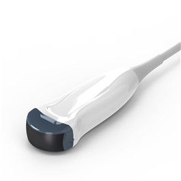
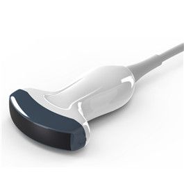
MC3-E
2.0 - 6.8 MHz Micro-Convex probe
MC6-E
4.0 - 12.0 MHz Micro-convex probe
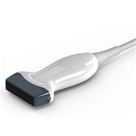
L7-E
4.0 - 15.0 MHz Linear probe
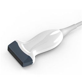
L12-E
7.0 - 18.0 MHz Linear probe
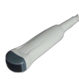
C3-E
2.0 - 6.8 MHz Convex probe

To install this Web App in your iPhone/iPad press ![]() and then Add to Home Screen.
and then Add to Home Screen.
